Figure 6.
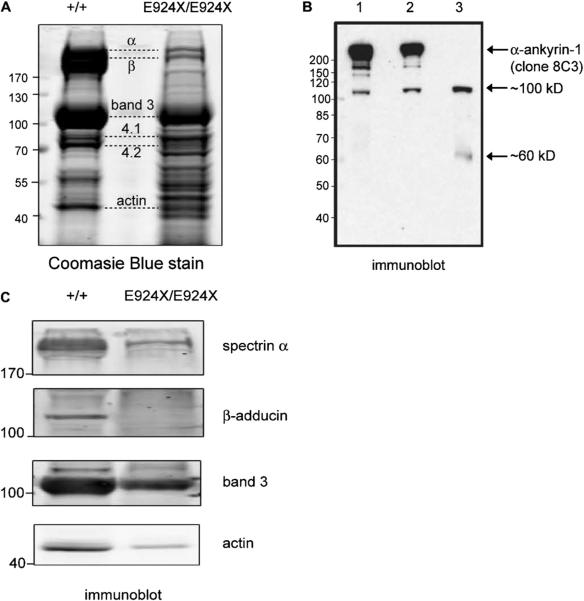
Several components of the band 3 macrocomplexes are destabilized in Ank1E924X homozygous mice. (A) Coomasie Blue stain of fractionated RBC ghosts (10% sodium dodecyl sulfate polyacrylamide gel electrophoresis) prepared from PB of WT (+/+) and homozygous Ank1E924X (E924X/E924X) mutant mice. Positions of the major RBC membrane proteins are indicated with dashed lines. (B) Immunoblot (α– ankyrin-1 monoclonal antibody [clone 8C3]) of fractionated RBC ghosts prepared from (1) WT (+/+), (2) E924X/+, and (3) E924X/E924X blood. Arrows indicate major immunoreactive bands. (C) Immuno-blot of fractionated RBC ghosts prepared from WT (+/+) and E924X/E924X blood probed with antibodies raised against major RBC membrane and cytoskeletal proteins. For all experiments, gel lanes were loaded with equal concentrations of total protein as measured by BCA assay. The size of molecular weight markers (in kD) and the identification of protein bands are indicated. Digital scans and LI-COR images were processed in Adobe Illustrator (11.0) with equal scaling and contrast enhancement, where required.

