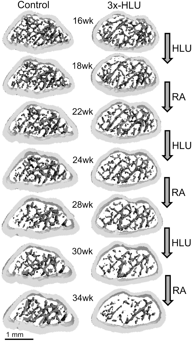
Fig. 2.
Volumetrically rendered three-dimensional morphology of the metaphyseal femur between 16 wk and 34 wk of age of a normally ambulating control mouse and a mouse that was subjected to three consecutive cycles of 2 wk HLU followed by 4 wk of RA. The consequences of superimposing HLU and RA onto the age-related decrease of trabecular number and increase in trabecular thickness are visible. For better visualization of individual trabeculae, only the central 25 (out of 74) slices of the region of interest are shown.

