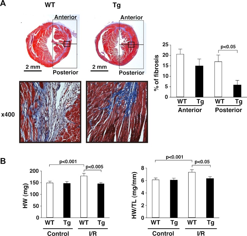
Fig. 2.
mTOR overexpression suppresses adverse LV remodeling. A: interstitial myocardial fibrosis. Left, representative photos of Masson's trichrome staining in cardiac sections from WT and mTOR-Tg mice at 28 days after I/R. Bottom, magnified images of the regions indicated by the squares in each heart section of the top images. Right, quantitative analysis of interstitial fibrosis examined by Masson's trichrome staining. n = 5 mice/group. B: heart weight (HW; left) and ratios of HW to tibia length (TL; right). Hearts from each group were harvested after echocardiography was performed. n = 11 control WT mice, 10 control mTOR-Tg mice, 11 I/R WT mice, and 16 I/R mTOR-Tg mice.

