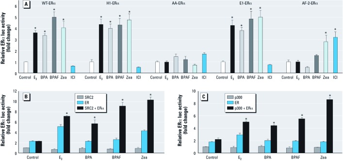
Figure 2.
Functional analysis of BPA, BPAF, and Zea on WT and ERα mutants and coactivation of ERα by SRC2 or p300 in Ishikawa cells. (A) For functional analysis, cells transfected with ERE-luc, pRL‑TK, and pcDNA/WT ERα, pcDNA/H1 ERα, pcDNA/AA ERα, pcDNA/E1 ERα, or pcDNA/AF2 ERα plasmid were treated with vehicle, 10 nM E2, 100 nM BPA, 100 nM BPAF, 100 nM Zea, or 100 nM ICI for 18 hr, and ERα-ERE–mediated activity was detected by luciferase reporter assay. Data shown are mean ± SE fold change relative to control for three independent experiments relative to control. (B,C) Coactivation of ERα by SRC2 (B) or p300 (C) in cells transfected with ERE-luc, pRL‑TK, and pcDNA/SRC2 or p300, pcDNA/WT ERα, or pcDNA/SRC2 or p300 plus pcDNA/WT ERα plasmids and treated with the vehicle, 10 nM E2, or 100 nM BPA, BPAF, or Zea for 18 hr. ERE-mediated activation was detected by luciferase reporter assay. Data shown are mean ± SE fold change relative to control for three independent experiments relative to control. *p < 0.05 compared with control. **p < 0.05 compared with the vehicle for co-transfections.

