Figure 3.
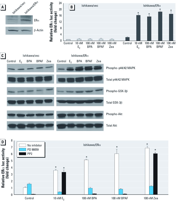
BPA, BPAF, and Zea affect p44/42 MAPK and src tyrosine kinase pathways in Ishikawa/ERα–stable cells. (A) Detection of ERα protein expression by Western blot in whole cell lysates prepared from Ishikawa/vec or Ishkawa/ERα cells. (B) ERE-mediated activity in cells transiently transfected with ERE-luc and pRL‑TK plasmids and treated with vehicle, 10 nM E2 or 100 nM BPA, BPAF, or Zea. Activity was detected by luciferase reporter assays, and data are mean ± SEM fold change relative to control for three independent experiments. (C) Western blot detection of phospho-p44/42 MAPK, phospho-GSK-3β, and phospho-Akt activation by 100 nM E2, 1,000 nM BPA, 1,000 nM BPAF, or 1,000 nM Zea. (D) Effect of PD 98059 and PP2 on the induction of PR gene expression by vehicle (control), 10 nM E2 or 100 nM BPA, BPAF, or Zea. PR transcripts were quantified by real time-PCR, and results are presented as mean ± SEM fold change relative to control for three independent experiments. *p < 0.05 compared with control.

