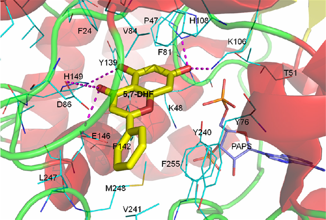
Figure 5. The docked pose of 5,7-DHF in SULT1A3.
5,7-DHF (yellow sticks) forms strong hydrogen bonding interactions (magenta dashed lines) with residue Asp86, Lys106, His108, and Glu146. In addition, hydrophobic interactions with Phe24, Phe81, Val84, Tyr139, Phe142, Tyr240 and Phe255 also stabilize the binding. The 7-OH is close to the catalytic residue His108 and orientated towards PAPS (light blue) for sulfonation. The ribbon diagram represents the secondary structure of SULT1A3.

