Figure 2.
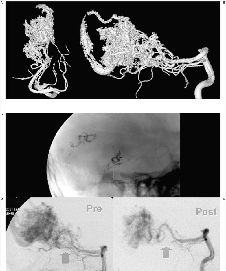
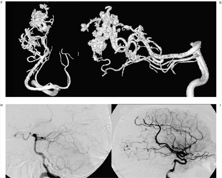
Case #14. LTS, 23-year-old man, clinical onset: haemorrhage. Splenial and Parasplenial AVM. A-B) 3D angiography from right vertebral artery. C) Onyx casts of two of the three pedicular Onyx injections, with targeted embolization of the AVM deeper portion (D-E), anatomically complex for surgical treatment. F-G) 3D angiography from right vertebral artery at the end of the last session. H-I) Left vertebral and right carotid angiography at the discharge, post surgery.


