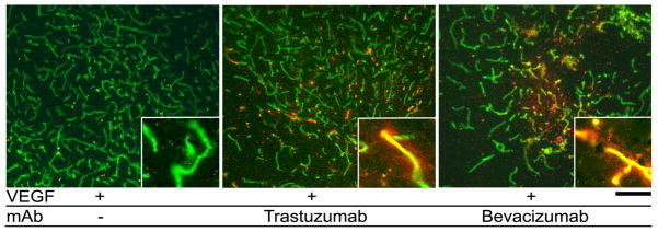
Figure 1.
Human antibody was detected in the brain parenchyma of mice treated with bevacizumab and trastuzumab. Vessels are labeled with lectin (green). Human IgG (red) was detected only in VEGF-stimulated regions of mice treated with bevacizumab and trastuzumab. Inserts show high magnification images of vessels. Scale bar: 100 μm.

