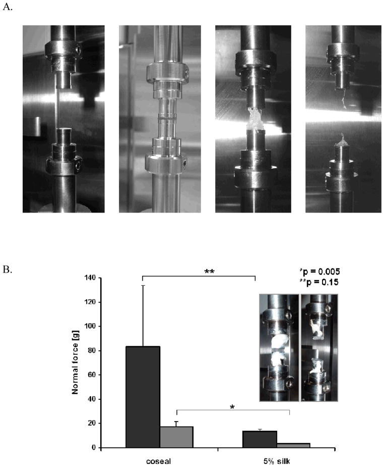
Figure 6.
A. Illustration of DMA measurement procedure (adhesion to steel) depicting the steel fixtures prior sample mounting (left) and the testing process. B. Comparison of CoSeal and 5% silk/PEG materials on intestines (dark grey) and steel (light grey), showing similar trends (n = 3). Inset – DMA setting for adhesion to intestines measurements. The indicated statistics were obtained with Student t-test.

