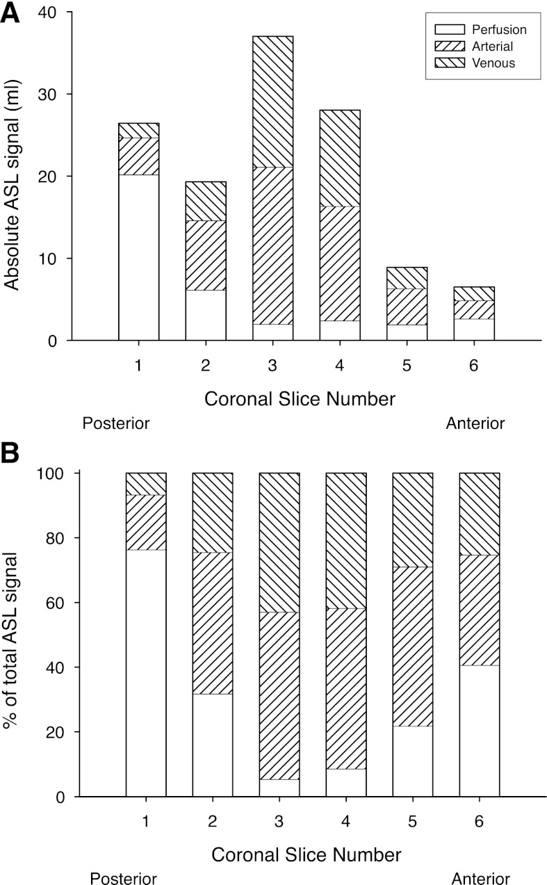
Fig. 4.
A: absolute ASL signal in each of the 6 coronal image planes studied. Location of the image planes are shown in Fig. 2B. B: fraction of the observed ASL signal (which necessarily adds to 100%) in each image plane resulting from the 3 blood compartments, arterial, venous, and true perfusion (blood delivered to or destined for capillary beds contained within the image plane).

