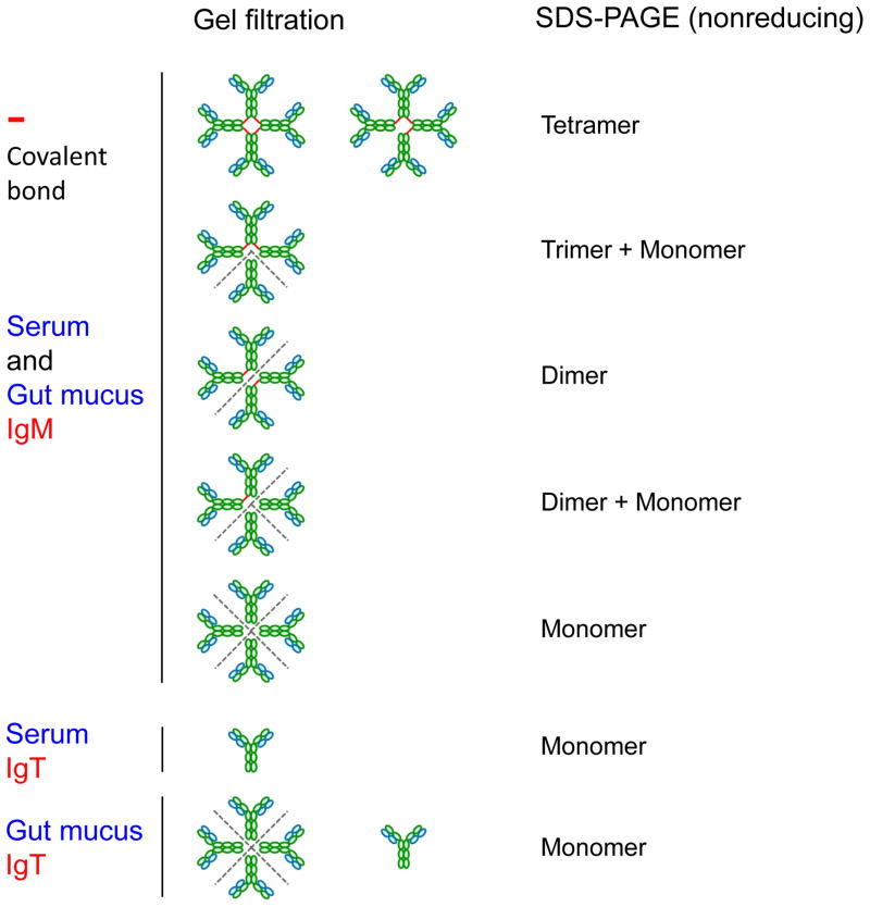
Fig. 2.
Redox forms of trout IgM and IgT in serum and gut mucus. Distinct chromatographic (gel filtration) and electrophoretic (nonreducing SDS-PAGE) behaviors of trout IgM and IgT are shown diagrammatically. Note the number and location of covalent bonds between the subunits of IgM are different in various redox forms [38]. The dotted lines indicate the areas where the non-covalent bonds are broken by SDS-PAGE under nonreducing conditions. The putative secretory component (SC) of a pIgR is not shown.

