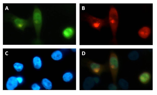
Figure 3.

PIM-1 and VDR co-localization in human keratinocytes. HaCaT cells were co-transfected with PIM-1-DsRed and VDR-YFP fusion constructs. (A) Image of the YFP-fluorescence of VDR. (B) Image of the red fluorescence of PIM-1. (C) Image of blue DAPI fluorescence. (D) Overlay of panels A, B and C showing colocalization of PIM-1 and VDR in the nucleus.
