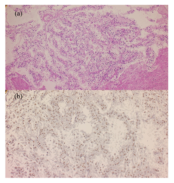
Figure 2.
Histological finding for left renal tumor. (a) Hematoxilin and eosin staining: the cytoplasm was clear or eosinophilic, and tumor cells proliferated with a papillary architecture or solid pattern; (b) transcription factor E3 immunohistochemical labeling of the left renal tumor: nuclei of many tumorous cells were diffusely positive for transcription factor E3.

