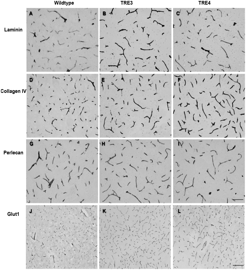
Figure 5. Levels and morphology of cerebrovascular basement membrane proteins in cortical capillaries of 3-month old wildtype, TRE3 and TRE4 mice.
Brain tissue sections from wildtype (a, d, g, j), TRE3 (b, e, h, k) and TRE4 mice (c, f, i, l) were processed for laminin, collagen IV and perlecan immunocytochemistry. The staining intensity of both laminin (a–c) and collagen IV (d–f) was higher in TRE3 and TRE4 mice, compared to wildtype animals. Perlecan levels were constant across genotypes (g–i). No differences were noted in the pattern of glut-l labeling of endothelial cells between mouse groups (j–l). Scale bars: a–l = 100 µm; m–o = 200 µm.

