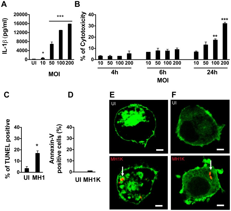
Figure 1. Burkholderia cenocepacia induces pyroptosis in macrophages.
Graphs in (A) through (D) represent mean ± SEM from three independent experiments. A. Macrophages were infected with B. cenocepacia MH1K at different MOIs for 1 h and then treated with gentamicin as described in Experimental Procedures. ELISA was used to quantify IL-1β in cell supernatants at 24 h post-infection. * p≤0.05 and *** p≤0.001 values compared to uninfected (UI) macrophages. B. Macrophages were infected as in (A) and the supernatants were used to quantify total lactate dehydrogenase (LDH) activity at 4, 6 and 24 h post-infection. ** p≤0.01 and *** p≤0.001 values for MOI of 10 at 24 h post-infection. C and D. Macrophages were infected as in (A) and at 24 h post-infection stained with the TUNEL-AF488 kit (C) or with Annexin V-AF488 (D) * p≤0.05 values compared to UI macrophages. Stained cells were analyzed by flow cytometry. E and F. Confocal images at 24 h post-transfection of macrophages expressing the fluorescent probe Lact-C2-GFP (Green) (E) or PH-PLCδ-GFP (Green) (F) Upper panels show UI macrophages. Lower panels show macrophages infected with B. cenocepacia MH1K-RFP (Red) for 4 h. Scale bar, 10 µm. Arrows indicate BcCVs.

