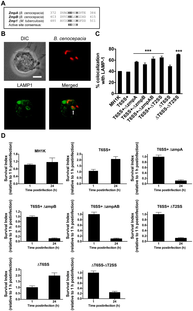
Figure 5. T2SS and its secreted substrates ZmpA and ZmpB are required to delay phagolysosomal fusion and for bacterial survival in macrophages in a T6SS-independent manner.
A. Alignment of the predicted ZmpA and ZmpB from B. cenocepacia with Zmp1 from Mycobacterium tuberculosis. The active site consensus is indicated in bold. B. Macrophages were infected with B. cenocepacia MH1K-RFP (Red) at an MOI of 50 for 4 h. Infected cells were fixed, permeabilized and stained with anti-LAMP-1 antibodies (green). Stained cells were analyzed by immunofluorescence microscopy. The arrow indicates LAMP-1 associated with the BcCV. Bar represents 10 µm. C. Macrophages were infected with B. cenocepacia MH1K, T6SS+, T6SS+-ΔzmpA, T6SS+-ΔzmpB, T6SS+-ΔzmpAB, T6SS+ΔT2SS, ΔT6SS or ΔT6SS-ΔT2SS, all expressing the red fluorescent protein (Red), for 1 h. Extracellular bacteria were removed by gentamicin treatment. Infected cells were fixed, permeabilized and stained with anti-LAMP1 antibodies (green). Quantification of LAMP1 associated with the BcCV is shown. Graph represents mean ± SEM from independent experiments including at least 60 vacuoles per experiment. *** p≤0.001 relative to LAMP1 associated to MH1K. (D) Macrophages were infected as in (C) and lysed at 1 and 24 h post-infection to quantify CFUs. The survival index at 24 h was calculated relative to the number of CFUs at 1 h post-infection. Graphs represent mean ± SEM from three independent experiments.

