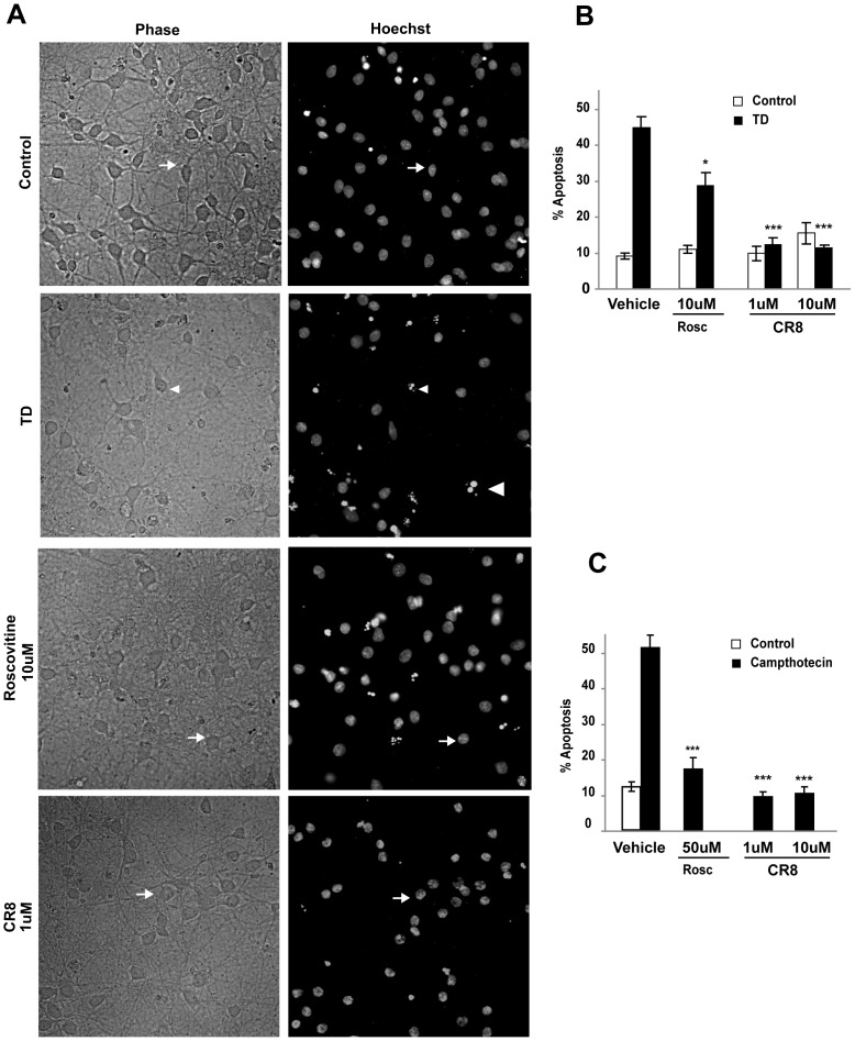
Figure 7. Pharmacological inhibition of CDK1 blocks neuronal apoptosis.
Cortical neurons were pre-treated with Roscovitine or CR8 (CDK1 inhibitors) or vehicle and then exposed to TD-or campthotecin induced apoptosis. A. Representative photomicrographs of control and trophic deprived neurons treated with the indicated concentrations Roscovitine and CR8 are shown. Upper row presents phase contrast images (Healthy neurons are indicated by larger cell bodies and abundant processes; Apoptotic neurons display shrunken cell bodies and sparse or lost processes). Lower row shows chromatin staining with Hoechst 33258. Arrows and arrowheads indicate surviving and apoptotic neurons, respectively suggesting an attenuation of TD-induced neuronal death in neurons pre-treated with Roscovinine or CR8. B. A quantitative assessment of the percentage of nuclei featuring chromatin condensation demonstrates a significant attenuation of TD-induced apoptosis in neurons pre-treated with Roscovitine (10 µM; *p<0.05, vs. TD vehicle) whereas CR8 at concentrations as low as 1 µM (***p<0.001, vs. TD vehicle) almost completely blocked development of apoptotic features in neuronal nuclei. C. Significant attenuation of campthotecin-induced apoptosis in neurons pre-treated with Roscovitine (50 µM; *p<0.001, vs. vehicle) and CR8 at concentrations as low as 1 µM (***p<0.001, vs. vehicle).

