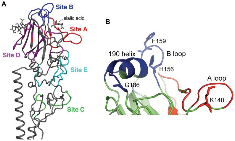
Figure 1. Antigenic structure of H3 HA.
Five antigenic sites A–E are mapped on the HA1 surface of H3N2 influenza viruses. (A). Antigenic site A (red color) and antigenic site B (blue color) are localized on the top of HA around the receptor binding pocket. (B). The “190 helix” and “B loop” create antigenic site B. The “A loop” is a part of antigenic site A. Figure 1A was made from PDB ID 2VIR [24] using PyMol (Schrödinger, LLC). Figure 1B was made from an Oklahoma/309 HA structural model made by SWISS-MODEL.

