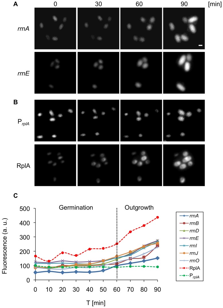
Figure 5. Expression of translational machinery components is biphasic during germination and outgrowth.
(A–B) Strains carrying PrrnA-gfp (AR13) (A-rrnA), PrrnE-gfp (AR16) (A-rrnE), PrplA-gfp (AR25) (B-PrplA), rplA-gfp (AR5) (B-RplA) were germinated in rich LB medium and tracked by time lapse fluorescence microscopy. GFP fluorescence images acquired at the indicated time points [min] are shown. Of note, the localization of RplA-GFP changed during germination from a diffuse dispersion pattern (0 min) into a distinct focus (30 min). Scale bar corresponds to 1 µm. (C) Spores of strains carrying Prrn-gfp (rrnO, A, B, D, E, I, J), PrplA-gfp or rplA-gfp were germinated in rich LB medium and tracked by time lapse fluorescence microscopy. GFP fluorescence images were acquired and analyzed at the indicated time points [min]. The data is representative of one out of three independent biological repeats. Fluorescence from at least 50 cells was measured and averaged for each time point and is shown in arbitrary units (a.u.) (see Materials and Methods). Scale bar corresponds to 1 µm.

