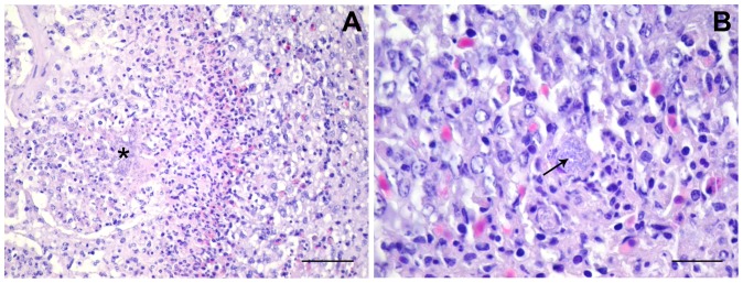
Figure 2. Necrotic foci in E. coli-infected lungs.
(A) UEL17-infected lungs. The boundary/edge of a necrotic focus containing extracellular bacteria (*). Scale bar 50 µm. (B) IMT5155-infected lungs. Arrow shows a macrophage with intracellular bacteria on the edge of a necrotic focus. Scale bar 20 µm.

