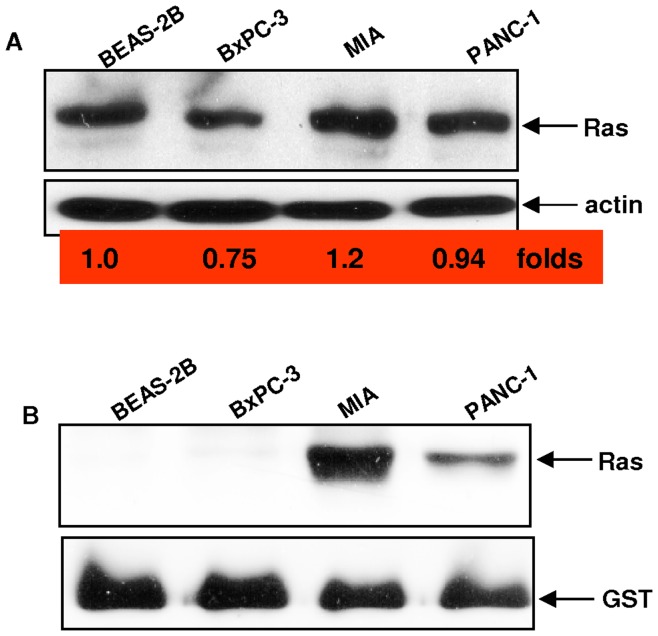
Figure 1. Activation status of Ras in pancreatic cancer cells.
A. The expression of Ras was examined by immunoblotting analysis in human pancreatic cancer BxPC-3, MIA, PANC-1 or lung epithelial BEAS-2B cells. The folds of the expression levels of Ras in pancreatic cancer cells relative to that in BEAS-2B cells were measured and indicated. Equal loading of total proteins per lane was determined by β-actin. B. Ras GTP-binding activity was measure in these cells by Ras-GTP assay. The blot was re-probed by anti-Ras antibody to judge evenly loading of total proteins.

