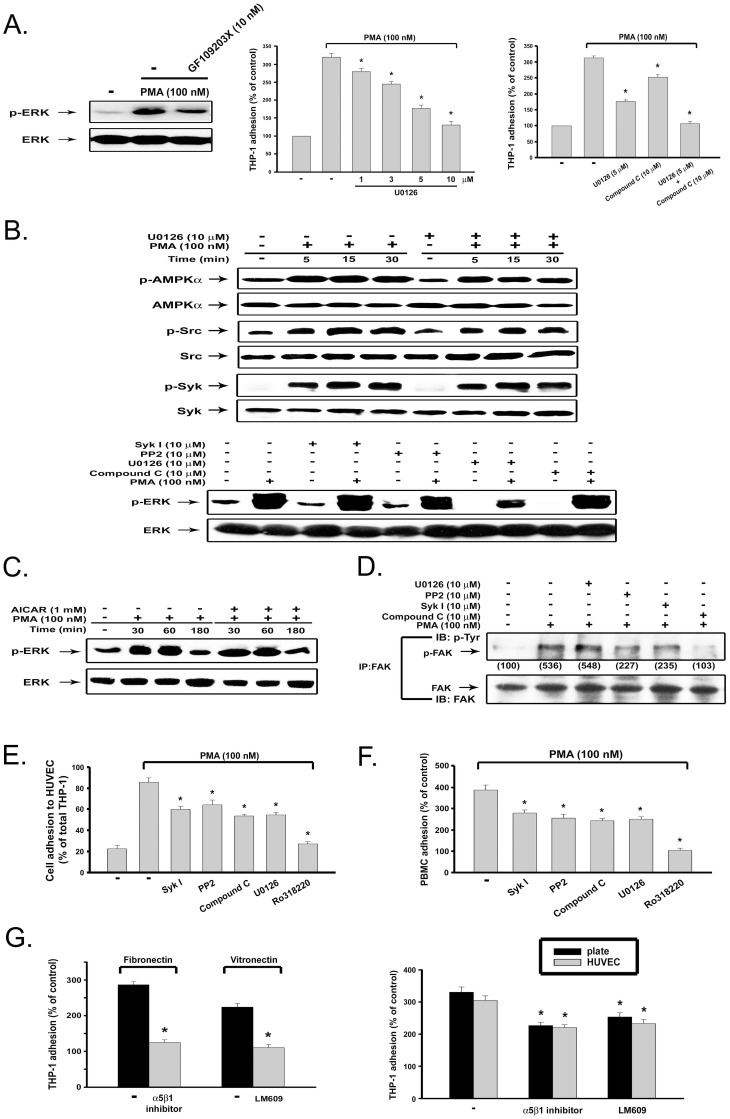
Figure 6. ERK activity independent of AMPK-Syk-Src signaling participates in PKC-mediated monocyte adhesion.
(A) After pretreatment with 10 nM GF109203X for 30 min, followed by PMA (100 nM) treatment for 10 min, cell lysates were subjected to immunobloting (left panel). In some experiments, after pretreatment with U0126 and/or compound C for 30 min, THP-1 cells were stimulated with PMA (100 nM) for 4 h, and cell adhesion was determined (middle and right panels). (B–D) THP-1 cells were treated with PMA in the presence of vehicle or pharmacological agents for 10 min (lower panel of B, D) or indicated time periods (upper panel of B, C). Immunobloting with specific antibodies was conducted. FAK activation was determined by immunoprecipitation with FAK-specific antibody and immunoblotting with phospho-tyrosine antibody (D). (E) THP-1 cells pre-labeled with BCECF were treated with SykI, PP2, compound C, U0126 (each at 10 µM), or Ro318220 (3 µM) for 30 min prior to the addition of PMA (100 nM). After 1 h incubation, THP-1 cells were washed with complete medium twice, and then added to HUVEC monolayer grown in 96-well plate. After 1 h, adhesion of THP-1 cells to HUVEC was determined. (F) Human primary monocytes were similarly treated with inhibitors and PMA, and cell adhesion at 4 h was determined. (G) PMA-induced cell adhesion to matrix-coated culture plates, in the absence or presence of alpha5beta1 inhibitor and alphaVbeta3 blocking antibody (LM609), was determined (left panel). In some experiments, after pretreatment with integrin inhibitors for 30 min, THP-1 cells were stimulated with PMA (100 nM) for 4 h, and cell adhesion to either the culture plate or to HUVEC was determined (right panel).

