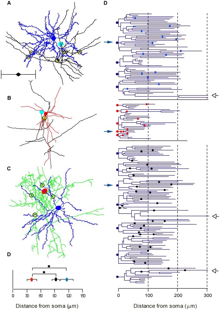
Figure 8. Location of LM contacts.
Contact sites seen at LM between the presynaptic axon and the postsynaptic dendrites were determined for all reconstructed pairs. A–C illustrates examples of an E–E, E–I and an I–E pair. The presynaptic soma of the spiny stellate cell is colored cyan and the axon is black. The basket cell’s soma and dendrites are colored red and the axon green. Postsynaptic spiny stellate cell is colored blue. For clarity, axons were trimmed to a sphere of about 400 µm diameter around the soma, but complete dendritic trees are presented. Putative contacts are marked by yellow-filled circles. Scale bar is 200 µm. D. Dendrograms of excitatory cells (somata indicated by blue circle) and inhibitory cells (somata indicated by red circle). Position of contacts on the dendrites are indicated by cyan (E–E) and red (E–I) rectangles. I–E contacts are indicated by black circles. Open arrowheads indicate three apical dendrites clipped at 300 µm E. The averaged distance of E–I contacts was significantly lower than E–E and I–E (P<0.05).

