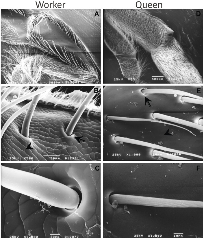
Figure 1. SEM images showing divergent morphologies of A. mellifera worker and queen hind legs during pupal development (Pb).
A: Worker hind leg external surface, note the bristle arrangement forming the pollen basket in the tibia, i.e., corbicula. B: Distal portion of the tibia of worker hind leg external surface. C: The single bristle on the worker hind leg external surface. This bristle may be a mechanoreceptor like the other bristles on the tibia of the worker hind legs. D: Queen hind leg external surface. E: Distal portion of the tibia of the queen hind leg external surface. F: A bristle on the queen hind leg external surface. Arrow points to bristle socket and arrowhead points to the structure of the cuticle. Original scale bars of scanning electron microscopy system.

