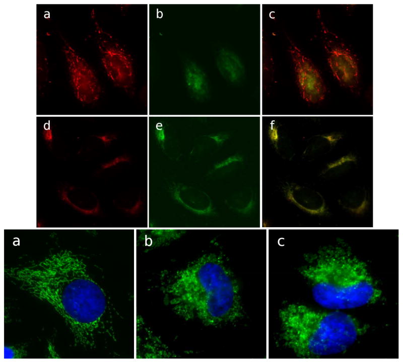
Figure 3.
Loss of mitochondrial membrane potential and structural integrity in cells treated with cluvenone. Mitochondrial membrane potential was examined in HeLa cells after treatment with 0.1% DMSO (a, b, c), or 1 μM cluvenone (d, e, f) for 1h, followed by staining with JC-1 for 15min. Cells with high membrane potential promote the formation of JC-1 aggregates which fluoresce red (a, d). In cells with low potential monomeric JC-1 fluoresces green (b, e). Yellow color in merged images (c, f) indicates depolarized mitochondria (Top Panel). Structural integrity of mitochondria was examined in HeLa cells treated for 4 h with 0.1% DMSO (a), or with 1 μM each of GA (b), or cluvenone (c). Fixed and permeabilized cells were then stained for the mitochondrial protein Tom 20 (green) as well as for nuclei, shown in blue (Bottom Panel).

