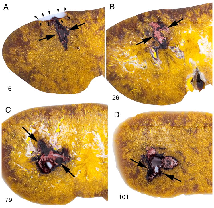
FIG. 2.
Path of lesion in pig kidney treated with the Storz SLX (2000 SWs, 120 SWs/min). These macroscopic images are of four sections (nos 6, 26, 79, 101) taken from 157 serial sections spanning the full thickness (anterior to posterior) of the kidney. The lesion can be tracked beginning at the posterior side (no. 6) where the parenchymal lesion (flanked by arrows) is continuous with an indentation caused by a subcapsular haematoma (blood washed out during processing). The lesion in sections 79 and 101 includes a region (arrowhead) in which haemorrhage was displaced during processing leaving a plug of embedding wax. This could only occur with complete ablation of tissue. In some cases such a channel could be tracked to the surface of the kidney. The lesion size for this kidney was determined to be 3.15% FRV.

