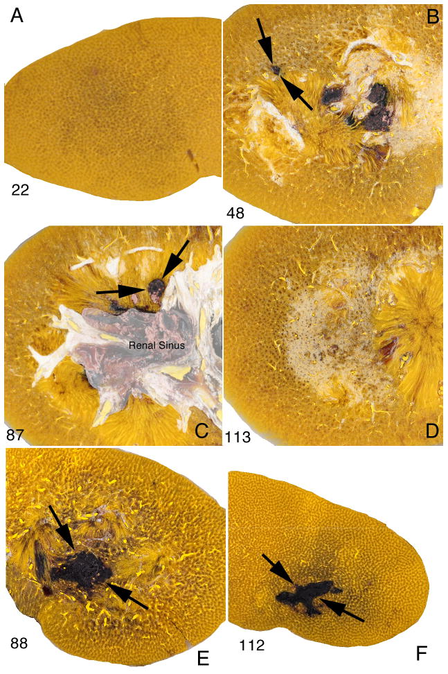
FIG. 3.
Comparison of lesion in pig kidneys treated at 60 SWs/min with Dornier HM3 and Storz SLX lithotriptors. Frames A–D show macroscopic images of four tissue sections (nos 22, 48, 87, 113) among 160 serial sections from a kidney treated at 60 SWs/min using the HM3 (2,000 SWs, 24 kV). The parenchymal lesion is limited to discrete regions in sections 48 and 87 (arrows). The lesion size in this kidney treated at slow SW rate measured 0.5% FRV compared with a mean value of 3.93% FRV for kidneys treated at 120 SWs/min as previously reported (Connors et al. [10]). Frames E and F show two sections (nos 88, 112) from a kidney likewise treated at 60 SWs/min but using the SLX (2000 SWs, PL-9). The lesion (flanked by arrows) is seen as a region of concentrated haemorrhage surrounded by seemingly unaffected parenchyma. The lesion volume in this kidney was 4.1% FRV.

