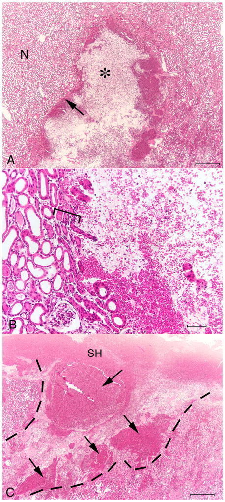
FIG. 4.
Histology of renal cortex from a pig kidney treated using the Storz SLX (2000 SWs, 120 SWs/min). (A) The haemorrhagic lesion (*) surrounded by intact kidney parenchyma. The portion of the lesion shown in this frame measures ≈ 2.5 mm × 3.0 mm. (B) The margin of the lesion, showing an abrupt transition between intact renal tubules and the core of the lesion largely devoid of organized structures. The transition from intact parenchyma to zone of complete tissue disruption occurs over a distance of just a few tubule diameters (square bracket). (C) Continuity can be seen between the parenchymal lesion (dashed line) and a subcapsular haematoma (SH). Several areas of intense haemorrhage are evident in the lesion (arrows). Bars, 0.5 mm (A), 0.05 mm (B), 0.5 mm (C).

