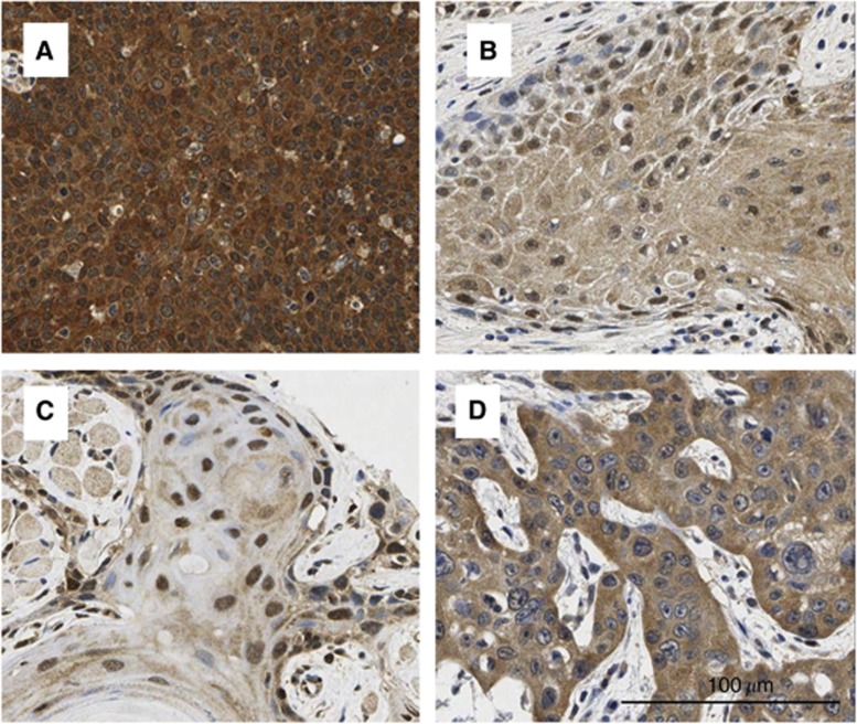
Figure 1.
Representative examples of p16 immunostaining in HNSCC. IHC staining for p16 expression of HNSCC was evaluated by product scores in different cellular compartments separately. From the above left: (A) p16 high expression in both the nuclei and cytoplasm; (B) p16 low expression in both the nuclei and cytoplasm; (C) high nuclear expression and modest cytoplasmic staining (however, by our scoring this still qualified at the lowest end of ‘high cytoplasmic’); and (D) high cytoplasmic expression and low nuclear expression.

