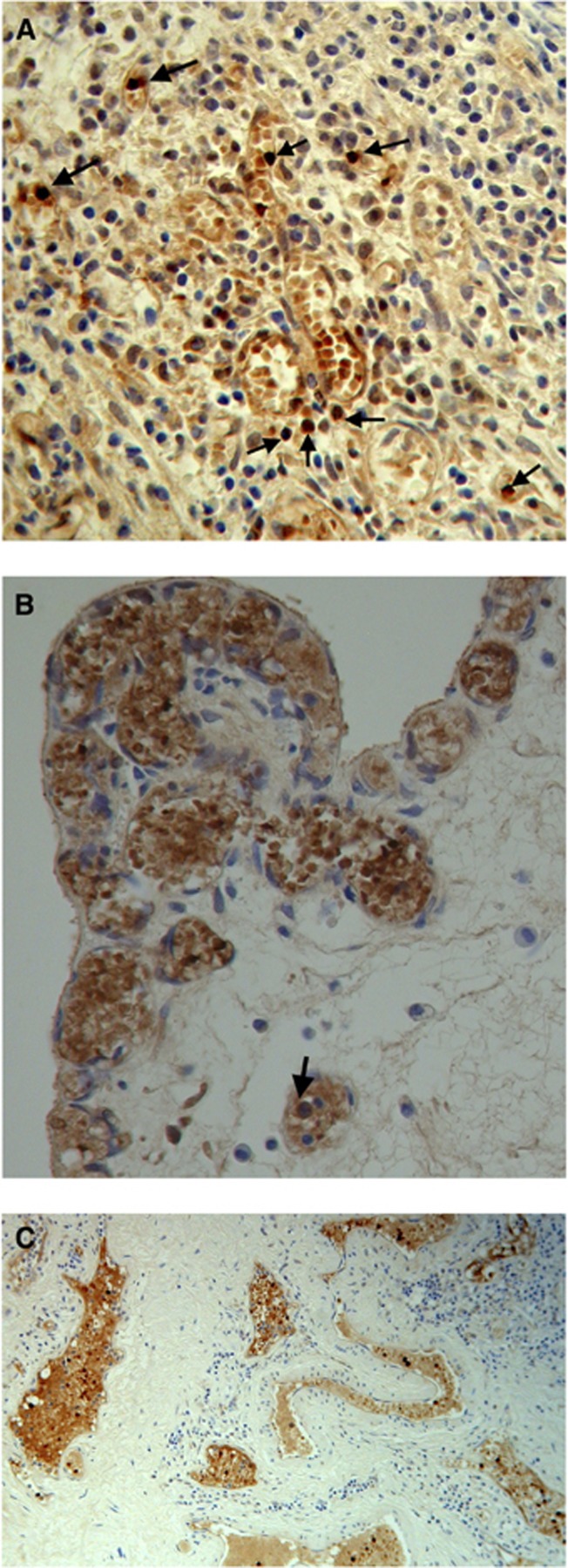
Figure 1.
Stages in proliferation of blood vessels with HbF cells in lamina propria of G1 TCC. Arrows indicating nucleated HbF progenitor cells: (A) clusters of HbF cells forming into fine vessels with many foci of nucleated HbF progenitor cells. (B) High density of small proliferating blood vessels. (C) Larger blood vessels spreading throughout the lamina propria.

