Figure 1.
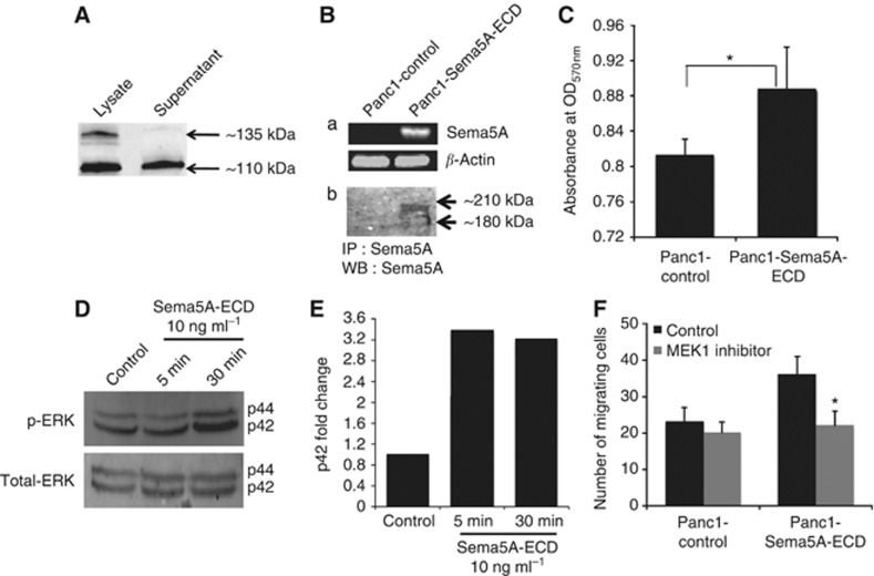
Secretion of SEMA5A and its invasive role in pancreatic cancer cells. (A) A representative western blot analysis showing expression of membrane-bound SEMA5A in protein lysates and secretion of SEMA5A in supernatant of human pancreatic cancer cell line – T3M4. (B) A standard (a) RT–PCR (using specific primers) and (b) immunoprecipitation and western blot analysis (using anti-SEMA5A antibody) of Panc1 cells ectopically expressing mouse Sema5A-ECD (Panc1-Sema5A-ECD) and control vector (Panc1-control) showing expression of Sema5A. β-Actin was used as an internal control for RT–PCR. (C) Cellular invasion in Panc1-Sema5A-ECD or Panc1-control after 1 h was measured using Matrigel-coated transwell chambers and MTT assay. Invasion was measured as mean optical density (OD) at 570 nm (OD570 nm)±standard deviation (s.d., bars) of triplicate culture and *a statistical significance of P<0.05. (D) Western blot analysis of phosphorylated and total ERK in protein lysates of Panc1 cells treated with 10 ng ml−1 of recombinant Sema5A-ECD protein at different time points (5 or 30 min) or media alone using appropriate antibodies. (E) Densitometric quantitation of phosphorylated p44 ERK that was normalised to total p44 ERK from western blots from (D) using the ImageQuant software. (F) Cellular migration in Panc1-Sema5A and Panc1-control after 4 h was measured after treating the cells either with 10 μℳ MEK1 inhibitor (PD98059) or media alone (control) using uncoated transwell chambers and the number of migrated cells was counted at × 10 magnification using a Nikon microscope. The values are number of migrated cells±s.d. (bars) of triplicate culture and *a statistical significance of P<0.05 using Student t-test.

