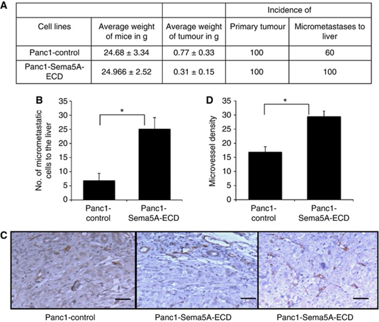
Figure 2.
Orthotopic assay, micrometastasis and microvessel density of Panc-1 Sema5A-ECD. Athymic nude mice (n=5) were injected orthotopically with Panc1-control or Panc1-Sema5A-ECD and were euthanised after 10 weeks. (A) Average weight of mice and tumour (tumour burden) as well as incidence of primary tumour and metastasis to the liver is shown. (B) There are a significantly enhanced number of micrometastatic cells per field of × 200 resolution of the Nikon microscope in the liver in Panc-1-Sema5A-ECD tumour-bearing mice as compared to control mice. (C) Immunohistochemistry using CD31 staining showing microvessels in Panc1-Sema5A-ECD and control orthotopic tumours. There is an increased angiogenesis in Panc1-Sema5A-ECD compared to the Panc1-control orthotopic tumours. (D) Densitometric quantitation of microvessel density from C. per field at × 200 resolution of the Nikon microscope. The values are number of microvessels±s.d. (bars) of five different areas per field or different slides and *a statistical significance of P<0.05.

