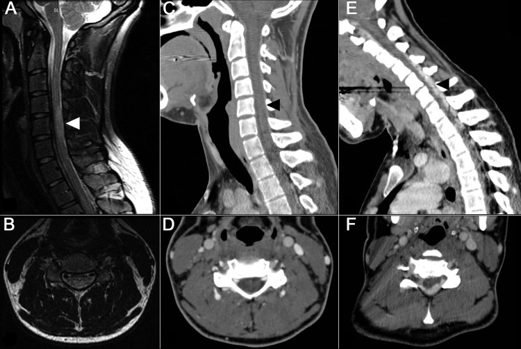
Figure. MRI and CT of the cervical spine.
(A) Sagittal and (B) axial T2-weighted MRI of the cervical spine in neutral position demonstrates central cord lesion at C4–C5 and T1–T2. (C) Sagittal and (D) axial CT venogram of the cervical spine in neutral position, at the level of C5. (E) Sagittal and (F) axial CT venogram of the cervical spine in flexed position, demonstrating enlargement of the epidural venous plexus, anterior displacement of the dura, and posterior compression of the cervical cord. Arrowheads on sagittal images indicate the spinal level of the corresponding axial image.

