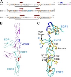
Figure 1.
Structure of 3 EGF domains of Del-1. A) Sequences of 3 EGF domains with secondary structures and disulfide bonds assigned. The linker between EGF1 and EGF2 is shown separately after EGF1. Brown arrow denotes β strands, and the blue wave sign indicates a short helix. Glycosylation sites are shown in red, and the RGD site in EGF2 is in cyan. The missing part of EGF1 in the current model is in italic and underlined with a broken line. B) The 2 molecules in ribbon drawing pack head-to-head in the asymmetric unit. The 3 EGF domains in the cyan molecule are labeled. The linker between EGF1 and EGF2 is in dark blue. C) α-Carbon skeleton drawing of the cyan molecule. View is rotated roughly 90° around a vertical axis from that in panel B. The RGD finger, the cation Ca, the metal-binding site, and 3 glycosylation sites are depicted. The 3 disulfide bonds in each of the EGF domains are shown in orange. The linker is in dark blue as in panel B.

