Figure 3.
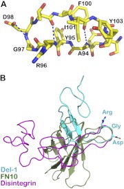
An RGD finger is located at the tip of a protruding loop. A) The RGD motif on a 13-residue loop of the Del-1 EGF2 domain. Note that Arg96-Gly-Asp-Thr form a β turn of the loop. One carboxyl oxygen atom of Asp98 forms a hydrogen bond with the main-chain nitrogen of the same Asp98 to stabilize the turn. Two main-chain hydrogen bonds and a few hydrophobic side-chain contacts help maintain the long loop. B) Superposition of RGD-containing loops from 3 proteins (superposition is based on Cα atoms of RGDX, X representing any other residues). EGF2 of Del-1 is in cyan, fibronectin type III domain 10 (PDB 1FNF) is in olive, and disintegrin (PDB 1J2L) is in magenta. The 3 proteins are conformationally very different, yet they all have their RGD positioned at the tip of a protruding loop.

