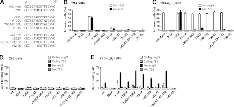
Figure 5.
Position of the Del-1 RGD sequence at the RGD fingertip is critical for interaction with integrin αVβ3. A) Point mutations or deletions in the RGD loop between C89 and C105 of Del-1. B, C) Adhesion of fluorescently labeled HEK293 control cells (B) or HEK293-αVβ3 transfectants (C) to Del-1-Fc proteins immobilized on protein A substrates. Adhesion was measured in buffer containing 2 mM Mn2+/0.2 mM Ca2+ and 25 μg/ml of either mouse IgG (open bars) or anti-β3 antibody 7E3 (solid bars). Adhesion to control substrates was <1.5% and subtracted from the data. D, E) Del-1-Fc proteins were complexed in solution with an FITC-labeled goat anti-human Fcγ antibody. Binding of multimeric Del-1 to HEK293 control cells (D) or HEK293-αVβ3 transfectants (E) in buffer with either 1 mM Ca2+/1 mM Mg2+ (open and striped bars), 2 mM Mn2+/0.2 mM Ca2+ (solid and shaded bars), in the presence of 25 μg/ml of either mouse IgG (open and solid bars) or anti-β3 antibody 7E3 (striped and shaded bars) was measured by flow cytometry. As a control, supernatant from mock-transfected cells was used for complex formation and gave mean fluorescence intensity (MFI) values of less than 8, which were subtracted from the Del-1 data to get specific binding. Human IgG1,κ control complexes bound with MFI values between 3.0 and 6.6. Data are averages ± sd from 2 independent experiments done either in triplicate (B, C) or in single determinations (D, E).

