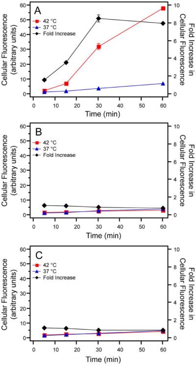
Figure 4.

Cellular uptake was quantified by measurement of fluorescence with live-cell flow cytometry. Fluorescence of cells incubated with Arg5-ELPBC at 42 °C increased significantly over the course of one hour, while those incubated at 37 °C showed little enhancement in fluorescence (A). ELPBC (B) and Arg5-ELP (C) controls showed minimal change in uptake over the course of an hour and showed little difference in uptake between 37 °C and 42 °C at each time point. The fold increase in cellular fluorescence (black line), calculated as the fluorescence at 42 °C divided by the fluorescence at 37 °C, reached approximately 8-fold for Arg5-ELPBC within one hour. Data represents the average of two replicates ± SEM.
