
Figure 7. Effect of the 1,25D3-MARRS neutralizing antibody on diosgenin-induced axonal regeneration.
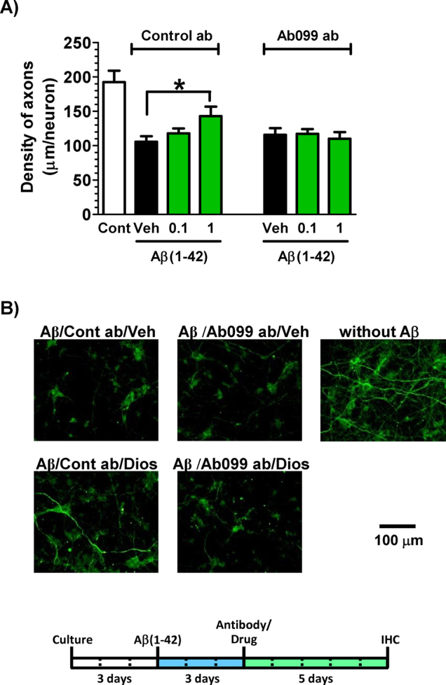
Cortical neurons were cultured for three days and then treated with or without aggregated Aβ(1–42) (5 μM). Three days after the administration of Aβ(1–42), the cells were treated with Ab099 antibody (Ab099 ab) or normal rabbit IgG (Control ab). After a ten-minute incubation period, diosgenin (0.1 and 1 μM) or vehicle solution (0.1% DMSO, Veh) was administered to the cells. Five days after treatment, the cells were fixed and double-immunostained for pNF-H and MAP2. The density of pNF-H-positive axons per MAP2-positive neuron was quantified for each treatment (A) (*p < 0.05, one-way ANOVA post hoc Dunnett's test, n = 7–9). (B) Representative images of each treatment are shown. The concentration of diosgenin was 1 μM.