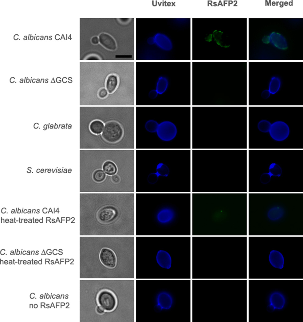
Fig. 3. Localization of RsAFP2 at the fungal cell surface.
Epifluorescence microscopy followed by deconvolution images of C. albicans (CAI4 and Δgcs strains), C. glabrata and S. cerevisiae treated with 50 µg/ml (native or heat-inactivated) RsAFP2 for 3 h and further incubated with Uvitex B2 (blue) and anti-RsAFP2 antibodies in combination with a FITC-labeled goat anti-rabbit IgG (green). Scale bars = 5 µm.

