
Fig. 1.
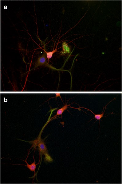
Perinatal asphyxia reduces neurite branching of primary cultured pyramidal neurons from hippocampus. Asphyxia was induced by immersing foetuses-containing uterine horns, removed from ready-to-deliver rats into a water bath at 37°C for 21 min. The cultures were prepared 6 h after delivery. After 14 days in vitro, the cultures were fixed with a formalin solution for assaying neuronal and astroglial phenotype using antibodies against microtubule associated protein-2 (MAP-2, red) and glial fibrillary acidic protein (GFAP, green) respectively, counterstained with 4′,6-diamidino-2-phenylindole (DAPI, blue), a DNA marker. A fluorescent photomicrograph of cultures from a caesarean-delivered control (a), and asphyxia-exposed (b) rats, showing MAP-2 (red) and GPAP (green) positive cells is shown. A significant decrease on neurite branching is observed in asphyctic cultures (b), principally evident in neurites of secondary and tertiary order. Moreover, a relationship between neurons and astrocytes can be observed in both experimental conditions, being more pronounced in astrocytes from asphyctic condition. Scale bar: 20 μm. Taken from Rojas-Mancilla et al., in preparation