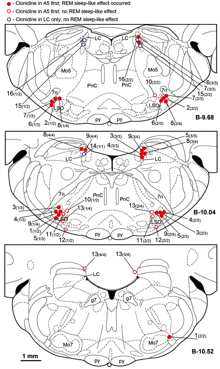
Figure 2.
Location of the sites of all clonidine injections made in this study. Each symbol represents the center of a separate injection site, as verified histologically from the location of Pontamine blue dye deposit (see Figure 1). Injection sites were superimposed onto the closest standard transverse brain section from a rat brain atlas (Paxinos and Watson, 1997) at the indicated levels measured from bregma (B). Each injection site is marked by the experiment (animal) number followed in parenthesis by the sequential order of the injection and the total number of sites injected in this experiment. Red filled and open circles correspond to the experiments in which clonidine was injected first into the A5 regions bilaterally and then, in some animals, also into the locus coeruleus (LC) uni- or bilaterally. Open blue circles correspond to the three experiments in which clonidine was injected into the LC only. Red filled circles indicate the experiments in which REM sleep-like episode occurred following clonidine injections; open symbols correspond to the experiments in which REM sleep-like episode did not occur at the end of the injection sequence (all three experiments with clonidine injections into the LC only and four experiments in which clonidine was first injected into the A5 region bilaterally but the injections were placed medial to the site where most noradrenergic A5 neurons are located). The injection sites illustrated in Figure 1 correspond to experiment 9 in this figure. Abbreviations: 7n, 7th cranial nerve; g7, genu of the facial nerve; Mo5, trigeminal motor nucleus; LC, locus coeruleus; LSO, lateral superior olivary nucleus; Mo7, facial motor nucleus; p, pyramidal tract; PnC, nucleus pontis caudalis.

