
Figure 5.
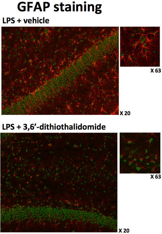
3,6′-Dithiothalidomide (56 mg/kg i.p.) treatment reduces the activation of astrocyte cells induced by the intercerebroventricular administration of LPS. Representative images of GFAP + ve cells in a field of view of the granule cell layer of the hippocampus are shown. The upper left panel (×20 objective magnification for both treatments) illustrates the highly activated numbers and morphology of astrocytes after the administration of LPS; the smaller image is a higher magnification of a section from the same image (×63 objective magnification, for both treatments). The lower panel illustrates how drug treatment markedly reduces the activated morphology of astrocytes after treatment with LPS; similarly to the above, this effect is further illustrated in the higher magnification side image. Astrocytes are stained red, whereas nuclei are counterstained green.