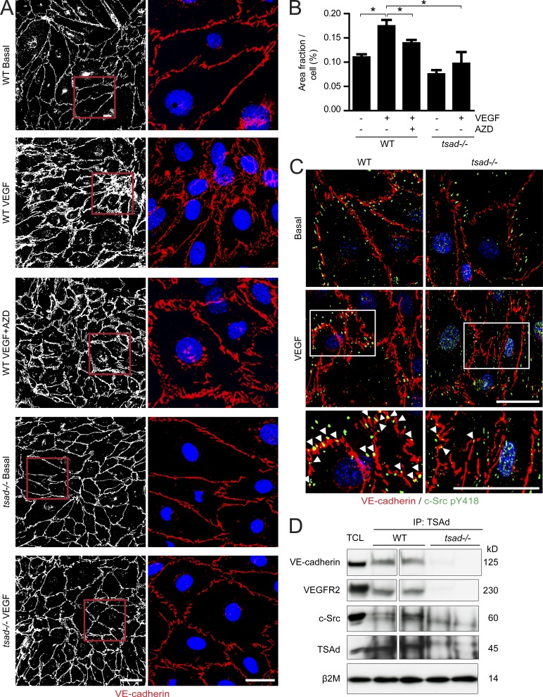
Figure 6.
Persistent adherens junctions in VEGF-stimulated isolated tsad−/− endothelial cells. (A) Primary endothelial cells isolated from WT or tsad−/− mouse lungs were cultured until confluent, and then treated for 10 min with vehicle or VEGF (100 ng/ml) with or without the Src kinase inhibitor AZD0530 (AZD; 10 µM). Cells were fixed and immunostained for VE-cadherin (left, white; right, red) and Hoechst 33342 (right, blue) to visualize adherens junctions and nuclei, respectively. Representative results from four separate cell isolations are shown. Bars, 50 µm (left row) and 20 µm (right row). (B) Quantification of VE-cadherin immunostaining in A, as VE-cadherin–positive area per cell, on 4 × 300 cells/genotype; 4 separate cell isolates. (C) Immunostaining for VE-cadherin (red) and c-Src pY418 (green) on isolated lung endothelial cells from WT and tsad−/− mice and treatment of confluent cultures for 10 min with 100 ng/ml VEGF. Bottom panels show magnification of boxed region in images from VEGF-treated samples. Arrowheads indicate c-Src pY418 localized in adherens junctions. Representative results from three separate experiments are shown. Bar, 20 µm. (D) Immunoprecipitation (IP) from lysates of WT and tsad−/− lung tissue using an anti-TSAd antibody, followed by immunoblotting for VE-cadherin, VEGFR2, and total c-Src protein. Total cell lysate (TCL) was used as a control to identify proteins in the IP and to control for equal protein concentration by immunoblotting for β2 microglobulin (β2m). Molecular weights in kilodaltons (kD) are indicated to the right. Data are representative of two independent experiments. n = 2 mice/genotype. Spacer line separates two parts of the same gel/blot, after excision of marker lane.

