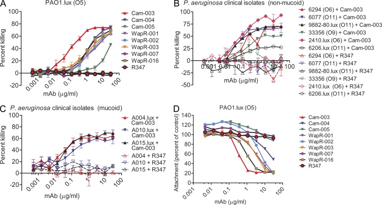
Figure 1.
Functional activity screening of antibodies derived from phage scFv patient libraries. In vitro functional screens included OPK assays and cell attachment assays using the lung epithelial cell line A549. R347, an isotype-matched human mAb that does not bind P. aeruginosa antigens, was used as a negative control. (A) Opsonophagocytosis assay with P. aeruginosa serogroup O5 strain PAO1, which was engineered to be luminescent (PAO1.lux), with dilutions of purified mAbs derived from phage panning. (B) Opsonophagocytosis assay with Cam-003 and heterologous serotype P. aeruginosa nonmucoid clinical isolates. Strains 9882–80, 2410, and 6206 were engineered to be luminescent (lux). (C) Opsonophagocytosis assay with mucoid P. aeruginosa clinical isolates that were engineered to be luminescent (lux). (A–C) The lines represent the mean percentage of killing and error bars represent the standard deviation. Percent killing was calculated relative to results obtained in assays run in the absence of antibody. (D) Log-phase PAO1.lux were added to a confluent monolayer of human cell line A549 cells after the addition of antibody at a MOI of 10 followed by analysis of RLUs after repeated washing to remove unbound P. aeruginosa. Wells lacking antibody were used as the comparative control. (A–D) Results are representative data from three independent experiments performed for each antibody and P. aeruginosa isolate.

