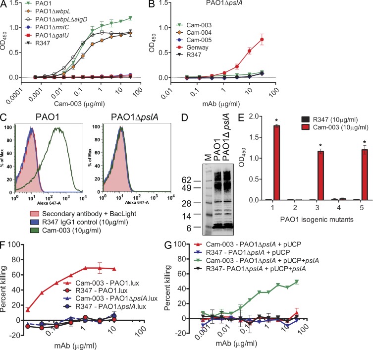
Figure 2.
Identification of the P. aeruginosa Psl exopolysaccharide as the target of antibodies derived from phenotypic screening. (A and B) Reactivity of antibodies was determined by whole-cell ELISA on plates coated with indicated P. aeruginosa strains: (A) wild-type PAO1, PAO1ΔwbpL, PAO1ΔwbpLΔalgD, PAO1ΔrmlC, and PAO1ΔgalU, and (B) PAO1ΔpslA. In B, mAb (Genway Biosciences) specific to a P. aeruginosa outer membrane protein was used as a positive control. (C) FACS binding analysis of Cam-003 to PAO1 and PAO1ΔpslA. Cam-003 is indicated by a green line; an isotype matched non–P. aeruginosa–specific human IgG1 antibody was used as a negative control and is indicated by the blue line. Washed cells were stained with BacLight to differentiate live from dead cells. Staining with the secondary antibody alone plus BacLight was used as an additional control. (D) LPS purified from PAO1 and PAO1ΔpslA was resolved by SDS-PAGE and immunoblotted with antisera derived from mice vaccinated with PAO1ΔwapRΔalgD, a double mutant strain deficient in O-antigen transport to the outer membrane and alginate production. (E) Cam-003 ELISA binding data with isogenic mutants of PAO1. Mutant 1, PAO1ΔwbpLΔalgD; mutant 2, PAO1ΔwbpLΔalgDΔpslA; mutant 3, PAO1ΔwbpLΔalgDΔpelA; mutant 4, PAO1ΔwbpLΔalgDΔpslA + pUCP; mutant 5, PAO1ΔwbpLΔalgDΔpslA + pUCP+pslA. * indicates P < 0.005 using the Mann-Whitney U test when comparing Cam-003 versus R347 binding. (F and G) Opsonophagocytosis assay using Cam-003 and negative control R347 with indicated luminescent (lux) strains of P. aeruginosa (F) or strains complemented with pUCP+pslA (G). R347 was used as a negative control in all experiments. (A–C, F, and G). (A–G) Results are representative data from three independent experiments.

