
Figure 3.
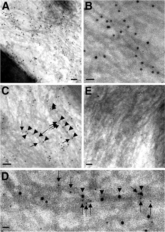
Ultrastructural colocalization of peripherin and NFL on the same NF in sciatic nerve by postembedding immunoelectron microscopy. Paraformaldehyde-fixed samples were incubated with rabbit anti-peripherin or mouse anti-NFL antibodies and probed with goat anti-rabbit IgG and goat anti-mouse IgG conjugated to 12 and 6 nm gold beads. As expected, for the immunodetection of peripherin in normal mice (A), large numbers of 12 nm gold particles are aligned with most 10 nm filaments in the axon. B, Higher magnification shows decoration of single filaments by 12 nm gold particles conjugated to anti-peripherin antibody. C, Linear arrays of two sizes of gold particles (12 nm for peripherin and 6 nm for NFL) decorate 10 nm filaments in the axon. D, Higher magnification shows that gold particles of two sizes overlie a single filament in the background. Arrowheads point to 12 nm particles (peripherin) and arrows to 6 nm ones (NFL). E, Negligible numbers of 12 nm gold particles (peripherin) are detected in peripherin knock-out mice, whereas 6 nm gold particles (NFL) are still present. Scale bars: A, 150 nm; B, 40 nm; C, 100 nm; D, 30 nm; E, 80 nm.