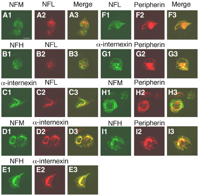
Figure 6.
Coassembly of peripherin with NF triplets and α-internexin into single filament network. SW13vim(−) cells were quintuple-transfected with constructs that expressed peripherin, NFL, NFM, NFH, and α-internexin and were immunostained with pairs of antibodies. Each set of three panels across represents the single immunolabels and the merged double label (yellow indicating colocalization) for the following pairs of antibodies: A1–A3, rabbit pAbs to NFL and mouse mAbs to NFM; B1–B3, rabbit pAb to NFL and mouse mAb to NFH; C1–C3, rabbit pAb to NFL and mouse mAb to α-internexin; D1–D3, rabbit pAb to NFM and mouse mAb to α-internexin; E1–E3, rabbit pAb to NFH and mouse mAb to α-internexin; F1–F3, rabbit pAb to NFL and mouse mAb to peripherin; G1–G3, rabbit pAb to α-internexin and mouse mAb to peripherin; H1–H3, rabbit pAb to NFM and mouse mAb to peripherin; I1–I3, rabbit pAb to NFH and mouse mAb to peripherin. Scale bar, 20 μm.

