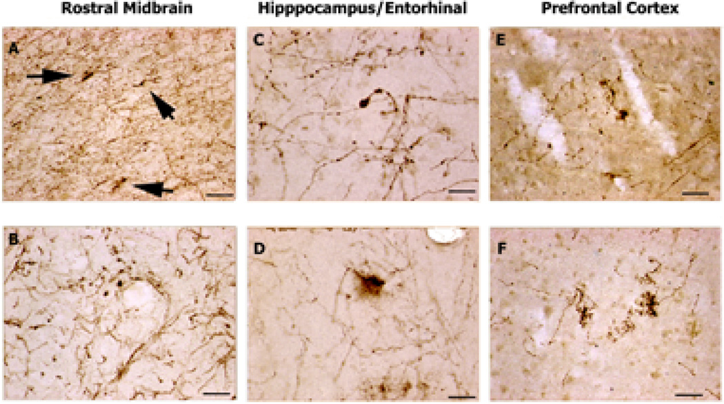
Figure 4. 5-HTT-IR Axons in Parkinson' Disease Donor Brain.
5-HTT-IR axons in brains from patients diagnosed with Parkinson’s disease. A. 5-HTT-IR axons in the red nucleus show some abnormal fibers (arrows) among many normal fibers. Scale bar is 200µm. B. In midline areas 5-HTT-IR fibers with enlarged varicosities are found among normal fibers. Scale bar is 50µm. C. Dystrophic 5-HTT-IR axons with enlarged varicosity in the dendritic regions of CA1. Scale bar is 50µm. D. Dystrophic 5-HTT-IR axons are splayed and degenerating in the upper layers of the entorhinal cortex. Scale bar is 50µm. E. Fine degenerating 5-HTT-IR axons in the upper layer of prefrontal cortex. Scale bar is 50µm. F. Fine, dense clusters of 5-HTT-IR axons in the deeper layers of prefrontal cortex. Scale bar is 50µm.

