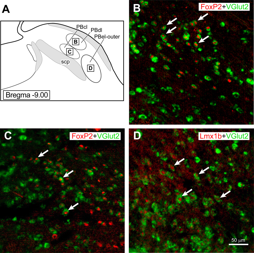
Figure 3.
(A) The parabrachial region showing the three PB subnuclei (PBdl, PBcl, and PBel-outer) with insert squares to indicate where photoimages presented in (B–D) were taken. Pseudocolors were added to the scanned images presents in B–C. (B): PBdl region that shows Vglut2 mRNA labeling in the neuronal cytoplasm (Gray et al.) and FoxP2 (red) immunoreactivity in the nuclei of these neurons. (B): PBcl region that shows Vglut2 mRNA labeling in the cytoplasm (Gray et al.) and FoxP2 (red) immunoreactivity. (C): PBel-outer region that shows VGlut2 mRNA labeling (Gray et al.) and Lmx1b immunoreactivity in the nuclei of these cells (red).

