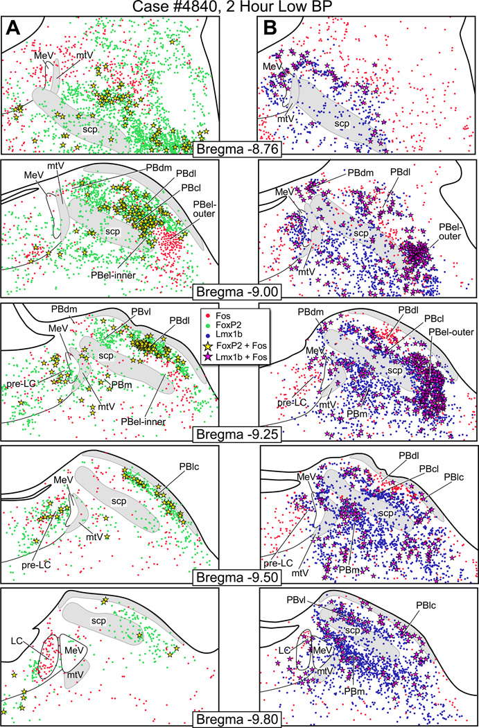
Figure 5.
Fos activation in the parabrachial nucleus after two hours of hypotension. (A) Line drawings showing Fos-activated neurons (red dots), FoxP2+ neurons (green dots), and co-labeled Fos & FoxP2+ neurons (yellow stars) after hypotension. (B) Fos-activated neurons (red), Lmx1b+ neurons (blue), and co-labeled Fos & Lmx1b+ neurons (magenta stars) after hypotension In Panel A, the bregma −9.0 shows a large concentration of Fos & FoxP2+ co-labeled neurons in the PBcl. In Panel B, the bregma −9.0 and −9.25 levels show a large number of Fos & Lmx1b+ co-labeled neurons in the PBel-outer.

